DIABETIC NEPHROPATHY: How is the treatment and prevention?


Treatment of Diabetic Nephropathy

The first step in treating diabetic nephropathy is treating diabetes and, if necessary, treatment of high blood pressure (hypertension). With your good blood sugar and hypertension management, you can prevent or delay renal dysfunction and other complications.


Drugs Diabetic Nephropathy

In the early stages of the disease, medication may be needed to:
• Control of high blood pressure. Drugs called angiotensin converting enzyme inhibitors (ACE) and angiotensin II receptor blockers (ARBs) are used to treat high blood pressure. Use of both drugs is not recommended because of the side effects that will arise. The results showed that blood pressure target was 140/90 millimeter mercury (mm Hg) or less.
• Management of blood sugar levels. Some medications have been shown to help control high blood sugar in people with diabetic nephropathy. The average target of hemoglobin A1C (HbA1C) is less than 7 percent.
• Lower high cholesterol. Cholesterol-lowering drugs called statins are used to treat high cholesterol and reduce protein in the urine.
• Improve bone health. Medications that help manage your calcium phosphate balance are important in maintaining bone health.
• Control of protein in urine. Drugs can often reduce the levels of protein albumin in the urine and improve kidney function.


Your doctor will recommend regular follow-up tests to see if your kidney disease remains stable or continues.

Treatment for advanced Diabetic Nephropathy disease

If your illness progresses to kidney failure (end-stage renal disease), your doctor will help you switch to a treatment focused on replacing your kidney function, with options including:
• Kidney dialysis This treatment is a way to get rid of toxic substances and dirt, and extra fluid from your blood. The two main types of dialysis are hemodialysis and peritoneal dialysis. The first method is more common and requires that you visit a dialysis center and connect to an artificial kidney machine about three times a week. Each session takes three to five hours. The second method can be done at home, but it requires compliance and discipline in the patient.
• Transplantation. In some situations, the best option is kidney transplant or kidney-pancreatic transplant. If you and your doctor decide on a transplant, you will be evaluated to determine if you are eligible for this surgery.
• Symptom management. If you choose not to have dialysis or kidney transplantation, your life expectancy is usually only a few months. You may receive treatment to help you stay comfortable.
Potential treatment of future Diabetic Nephropathy
In the future, people with diabetes nephropathy can benefit from the treatment being developed by using regenerative medicine. This technique can help restore or slow down kidney damage due to this disease. For example, some researchers think that if a person's diabetes can be cured with future treatment such as pancreatic pancreatic cell transplantation or stem cell therapy, then renal function may improve.
In addition, studies on stem cells and some new drugs for Diabetic Nephropathy are being developed.


 Lifestyle and treatment of Diabetic Nephropathy at home
• Lifestyle behavior can support treatment. Active in physical activity every day of the week, with at least 30 minutes of physical activity every day of the week.
• Adjust your diet. Talk with a dietitian about limiting salt intake, choosing foods that have lower potassium levels and limit the amount of protein you eat.
•          Quit smoking. If you are a smoker, talk to your doctor about strategies to quit smoking.
• Maintain a healthy weight. If you need to lose weight, talk to your doctor about weight loss strategies. Often this involves an increase in daily physical activity and reduced calories.
• Take aspirin every day. Talk to your doctor about whether low-dose aspirin every day is useful to you.


Support Against Diabetic Nephropathy Patients

If you are susceptible to diabetic nephropathy, here are some steps that may help you to overcome it:
• Connect with others who have diabetes and kidney disease. Ask your doctor about support groups in your area
• Maintain your normal routine, whenever possible. Try to keep the normal routine, do the activities you enjoy and continue to work, if conditions are possible. This can help you cope with the sadness or loss you may experience after a diagnosis of the disease.
• Talk to someone you trust. Living with diabetic kidney disease can create stress, and by telling stories, can help your feelings. You may have friends or family members who are good listeners. Or you may find it helpful to talk to a faith leader or someone you trust. Consider asking a doctor's referral to a social worker or counselor.


Prevention of Diabetic Nephropathy
To reduce the risk of kidney disease diabetes:
• Control diabetes. With effective diabetes treatment, you can prevent or delay diabetic kidney disease.
• Control of high blood pressure or other medical conditions. If you have high blood pressure or other conditions that increase your risk of kidney disease, go to your doctor. Ask your doctor about the examination to look for signs of kidney damage.
• Follow the instructions on over-the-counter medications. For people with diabetic kidney disease, taking this type of painkiller can cause kidney damage.
• Maintain a healthy weight. If you have a healthy weight, try to keep it physically active every day of the week. If you need to lose weight, talk to your doctor about weight loss strategies. Often this involves an increase in daily physical activity and reduced calories.

• Avoid smoking. Smoking can damage your kidneys and make your kidney damage worse. If you are a smoker, talk to your doctor about smoking cessation strategies. Counseling and treatment can help you quit smoking.

Diabetic Nephropathy: What is the cause and who is at risk?


Causes of Diabetic Nephropathy

Diabetic nephropathy occurs when diabetes disease damages blood vessels and other cells in your kidneys.

How the kidneys work

Your kidneys contain millions of tiny blood vessels (glomeruli) that filter out toxins and impurities from your blood. Severe damage to these blood vessels can cause Diabetic Nephropathy, decreased renal function and kidney failure.

Causes of Diabetic Nephropathy

Diabetic nephropathy is a common complication of diabetes, in type 1 and 2 diabetes.

Over time high blood sugar, associated with uncontrolled or untreated diabetes can cause high blood pressure. Furthermore, this will damage the kidneys by increasing the pressure on the kidney filtering system (glomeruli)


Risk factors Diabetic Nephropathy

Several factors may increase your Diabetic Nephropathy risk, including:
• Diabetes, type 1 or 2
• High blood sugar (hyperglycemia)
• High blood pressure (hypertension)
• Smokers and diabetics
• High blood cholesterol levels and diabetes
• Family history of diabetes and kidney disease


Complications of Diabetic Nephropathy

Diabetic Nephropathy Complications can occur gradually over months or years. These include:

• Fluid retention, which can cause swelling in the arms and legs, high blood pressure, or fluid in your lungs (pulmonary edema)
• Increase in potassium levels in your blood (hyperkalemia)
• Heart and blood vessel disease (cardiovascular disease), may lead to stroke
• Damage to the blood vessels of the eye (diabetes retinopathy)
• Anemia (lack of blood)
• Foot wounds, erectile dysfunction, diarrhea and other problems associated with damaged nerves and blood vessels
• Complications of pregnancy that pose a risk to the developing mother and fetus
• Irreversible damage to your kidneys (end-stage renal disease), which ultimately requires dialysis or kidney transplantation to survive.


Diagnosis of Diabetic Nephropathy

If there are signs and symptoms, as well as the above-mentioned risk factors, you can check with your family doctor. Physical examination, and interviews pertaining to your medical history are required to determine the next course of action. Your family doctor may refer you to a specialist kidney (nephrologist) or diabetes specialist (endocrinologist).


To find out if you have diabetes kidney disease, you may need certain tests and procedures, such as:
• Blood tests. If you have diabetes, you need a blood test to monitor your condition and determine how well your kidneys work.
•          Urine test. The urine sample provides information about your kidney function and whether you have too much protein in the urine. A high level of protein called microalbumin may indicate that your kidney is exposed to diabetic kidney disease.
• Imaging tests. Your doctor may use X-rays and ultrasound to assess the structure and size of your kidneys. You may also have CT scans and magnetic resonance imaging (MRI) to determine how well the blood circulates in your kidneys. Other imaging tests can be used in some cases.
• Kidney function testing. Your doctor can assess your kidney screening capacity by using renal analysis analysis.
• Kidney biopsy. Renal tissue retrieval may be necessary for further examination of the microscopic renal structure.

How is the treatment of this disease?

DIABETIC NEFROPATI


WHAT IS THAT??

Diabetic nephropathy is a renal-related complication in type 1 diabetes and type 2 diabetes. It is also called diabetic kidney disease. As many as 40 percent of people with diabetes will develop kidney disease.

Diabetic nephropathy affects the kidneys' ability to perform functions in removing toxic substances, and excess fluids from your body. The best way to prevent or delay Diabetic Nephropathy is by maintaining a healthy lifestyle and treating diabetes and high blood pressure.

Over the years, diabetes is slowly destroying your kidney's difficult-to-do screening system. Early treatment can prevent or slow the progression of the disease and reduce the likelihood of complications.

Kidney disease can develop into kidney failure, also called end-stage renal disease. Kidney failure is a life-threatening condition. At this stage your treatment option is dialysis or kidney transplantation.


Symptoms and signs of Diabetic Nephropathy

In the early stages of Diabetic Nephropathy, you may not notice any signs or symptoms. In the next stage, signs and symptoms that arise include:

• Uncontrolled blood pressure
• The presence of protein in the urine
• Swelling of the feet, ankles, hands or eyes
• Urinate a lot
• Reduced drug or insulin requirement to treat diabetes
• Confusion or difficulty concentrating
• Loss of appetite
• Nausea and vomiting
• Itching in the skin
• Easily tired

When to see a doctor?

Make an appointment with your doctor if you have signs or symptoms of kidney disease.

If you have diabetes, Urine examination to detect protein needs to be done every year. It helps determine how well the kidneys function.

What causes this disease?

Dengue Hemorrhagic Fever Management


Dengue Hemorrhagic Fever (DHF) is a form of dengue infection accompanied by bleeding manifestation from mild to severe. DHF becomes a health problem both in tropical and sub-tropical areas (as "imported cases", cases under the tropics).

Since the advent of the disease several decades ago, to date there has practically no decrease in both incidence and prevalence. Dengue fever is a fever caused by Dengue virus infection.

Known 4 different types of Dengue Virus namely: Den-1, Den-2.Den-3, and Den-4.

Clinical signs and symptoms

Usually asymptomatic (no symptoms). Patients taken to the hospital are usually in severe condition. Symptoms and signs-clinicnya syndrome (collection of symptoms) from mild form of mild fever to shock due to severe bleeding.

Bleeding and plasma leakage (leakage of blood vessels) that occur usually because the patient gets an infection attack by one type of Dengue virus. In endemic areas (like Indonesia) we should suspect any fever that occurs as dengue fever until further examination proves that it is not dengue fever.

Specialist surgeons (who suspect there is appendicitis) and obstetricians (who suspect there is an out-of-date pregnancy) should be vigilant when confronted with patients from endemic areas or recently returned from endemic areas with high fever accompanied by abdominal pain (abdomen ).

Do not rush into surgery, before being sure that the stomach pain is not due to dengue fever. Because the symptoms are similar between dengue fever with appendicitis, and pregnancy outside the womb (ectopic pregnancy is disrupted).

Laboratory examination is characterized by decreased platelets and signs of plasma Leakage (inflammation of the plasma due to leakage of blood vessels) is the occurrence of hemokonsentrasi (increased hematocrit levels). Clinical and laboratory changes of DHF patients Very quickly, requiring strict vital monitoring and serial laboratory (every 12 hours or 24 hours).

WHO criteria are still used for the diagnosis of DHF. But in certain circumstances we should not only hold on to one kind of examination. Platelet value less than 100,000 (only) can not be used as a handle to insert the patient to the hospital, because platelets that are still above 100,000 / mm3 can suddenly drop drastically. So if the patient is still at home would be very dangerous to the soul of the patient.

Diagnosis of DHF with serological examination of ELISA and rapid test (rapid test) with Dengue blot. For Dengue Blot usually new positive on fifth day of fever. There are other checks: NS1 antigen, recommended for patients with fever less than 3 days, if positive supports Dengue Fever, But if negative results should be confirmed again with Dengue Blot after fever day 5.

Management

Dengue Hemorrhagic Therapy is supportive (boosting the immune system) and symptomatic (relieving symptoms). No specific drugs have been found to kill the Dengue virus.

Need to replace fluid loss due to plasma leakage (Dengue virus attacks blood vessel walls) and provide substitution therapy (replacement) of blood components when needed. If platelet counts are very low and bleeding occurs, platelet transfusions are given.

In the administration of fluid therapy, the need for fluid monitoring. Take care of patients both clinically and laboratories (see Hemoglobin, Hematocrit, and platelet levels). The process of plasma leakage and the occurrence of thrombocytopenia (decreased thrombocyte) generally occur days 4 to 6 since the fever. Thus, it is necessary to be careful when treating DHF on days 4 to 6. On that day patients often do not complain of heat and tend to ask for outpatient care.

Day 7 fever, the process of plasma leakage will decrease and the fluid returns from the interstitial space (around blood vessels) to the intravascular (into the blood vessels). Liquid therapy in such circumstances should be gradually reduced. Because, will cause a lot of fluid buildup in the blood vessels.

It is necessary to monitor the possibility of fluid overload and the occurrence of pleural effusion (accumulation of fluid in the lung layer) or ascites (fluid accumulation in the abdominal cavity). Can be seen from clinical symptoms: shortness of breath, heavy breathing, and feelings of discomfort.

Need nonfarmakologis Therapy (without drugs) which includes bed rest (on platelets openia = severe platelet count). Normal platelet levels: 150 thousand to 450 thousand. If dropped below 100 thousand, should be hospitalized. Because it is feared there will be bleeding and possible Shock (Shock Syndrome in Dengue).

Provision of food with nutritional content: regular rice or soft rice according to the taste of the patient. Needed a meal Does not contain substances or spices that irritate the digestibility (spicy, sour).


 Symptomatic therapy, given antipyretics (paracetamol), overcomes dyspepsia (discomfort in the pit of the stomach, nausea, vomiting, sebah, easily satiated, bloated, often belching).

The fluid administration protocol as a major component of adult DHF management follows 5 protocols, which refer to the WHO protocol.

The protocol is divided into 5 categories, as follows:
1. Handling suspect dengue without shock.
2. Giving fluids to adult DHF suspects in the care room.
3. Management of DHF with increased hematocrit (blood viscosity)> 20%
4. Management of spontaneous bleeding in adult dengue
5. Management of dengue shock syndrome in adults

Dengue Hemorrhagic Fever will always be there throughout the year. We can prevent transmission of this disease by jointly seeking the breaking of the transmission chain by eradicating mosquito nests.

By: Dr. Muchlis Achsan Udji Sofro SpPD-KPTI

SMF Disease In RSUP Dr Kariadi Semarang

RECIPIENT TREATMENT OF TYPHOID DISEASE


Typhoid disease (Typhoid Fever) is still a global problem today. Not only in Indonesia, worldwide Tifes disease is still a community problem. The problems that arise are in terms of how to make the diagnosis, how to handle, and how to prevent it.

To make a diagnosis for example, doctors sometimes have difficulty remembering symptoms often resemble other infectious diseases, such as: Dengue Fever, Leptospirosis, Malaria, Urinary Tract Infection, or Breathing Infections.

To ensure Diagnosispun, not all hospitals are able to perform blood culture examination. As a result, only with clinical symptoms and a simple investigation (Widal's test) is diagnosed.

The selected drug (first choice) for Tifes treatment is Chloramphenicol. Most doctors still use the drug. Now begin to emerge a new problem that is: the recurrence. Not a few patients who get tifes treatment is easy to relapse.

Furthermore the problem of drug resistance. Germs that cause Tifes, Salmonella typhi, some have resistance to chloramphenicol drugs. That is, if given the drug, then the germs are immune to natural mechanisms.

Symptoms and signs

Incubation period (inclusion of germs until symptoms develop) about 10-14 days (5-23 days). Symptoms include: acute fever (sudden), fever chills, headaches and disturbances around the stomach (nausea, vomiting, discomfort in the uluhati) and decreased appetite.

Signs of Typhoid disease:
Fever rises gradually, sometimes up to 41 ° C. This temperature rise can take place continuously, or up and down. Generally body temperature will increase in the afternoon. There is a relative bradycardia, ie the increase in pulse rate is not proportional to the rise in body temperature.

There is a tifes tongue in the form of: reddish tongue with white tongue edge. Some patients have meteorismus: a bulging abdomen due to weak bowel movement, so air is retained in the intestine and difficult to remove. Sometimes patients experience enlarged liver and spleen (hepatosplenomegaly).

Diagnosis

To establish a diagnosis of Tifes, can still use the Typhoid Fever Clinic Score. This score gives the value of one and two of each symptoms and signs experienced by the patient. A single score is given for symptoms: fever less than a week, headache, body "nglungrah" (not powerful), nausea, abdominal pain and decreased appetite. One score is also given for signs: vomiting and decreased bowel movements.

While the two scores are given for: difficulty sleeping, enlarged liver and spleen, and fever that is more than one week.

When added, the clinical score of Tifes can be up to 20. However, if the clinical score reaches 13 or more it can be stated that Clinical Typhoid Fever.
In order to help establish the diagnosis of Tifes, laboratory investigation is required. Among other blood routine occasionally found leukopeni (decreased leukocyte count).

Widal laboratory tests can still be used, but should be more careful in providing interpretations of Widal. In some patients Widal positive results will last for months, some even up to one year after the attacks Tifes still positive. But that does not mean it is now attacked by Tifes.

A subsequent examination that is helpful in establishing the diagnosis of Tifes is IgM Salmonella (Tubex TF). This check produces positive results in the heat of the fourth or fifth day. And some will Survive to remain positive for up to 35 days or there are up to two months still positive.

Therefore, do not protest to the doctor if it is healed from tifes kok results IgM Salmonelanya still positive. This is our immune to the germs of tifes. Not that we're tifes, or relapse tifesnya, as long as there is no fever, although IgM Salmonelanya positive.
So, we should use the results of laboratory tests as a support for the diagnosis. Do not treat laboratory results if there are no symptoms and signs that lead to typhoid fever (Tifes).

Recent Handling

Handling Tifes is not enough with antibiotics. Need general care and not always hospitalized. Indications are hospitalized if the patient vomits repeatedly, high fever that does not come down with the medicine down the heat, and the body is not weakened.

Antibiotics are the main choice until now is still chloramphenicol group. However, the weakness of this drug should be given for 14 days and a day three to four times drinking. Though some patients are often the seventh day is not fever and the drug will soon be stopped by itself. As a result the treatment becomes ineffective and allows for relapse again. In addition it will facilitate the resistance of germs (immune) to the antibotics given.

The next antibiotic is the Fluoroquinolone group Ciprofloxacin and Levofloxacin. Ciprofloxocin is a good antibiotic for Tifes. This drug is able to pursue the germs that cause tifes to the bone marrow (where hiding Salmonella Typhi in our bodies). Only this drug is given within seven days with a dose twice a day.

Recent antibiotics that have been studied in several cities in Indonesia (Jakarta, Bandung, Semarang, Malang, Denpasar, Makassar) are Levofloxacin. The advantage of this drug, able to reduce heat earlier than ciprofloxacin. In addition, the side effects (nausea, vomiting, abdominal discomfort, impaired liver function) are lighter than ciprofloxacin. And given for seven days but with a sufficient dose once a day. For patients it would be more convenient if only taking medication once Day (compare with chloramphenicol 3-4 times, ciprofloxacin 2x daily).

Levofloxacine antibiotics are known for respiratory tract infections, lung infections (pneumonia) and urinary tract infections. With the research was able to overcome the germ Salmonella Typhi with good results.

Now, many researchers have shown that early dense feeding of rice with low-cellulose side dishes (abstinence vegetables with coarse fiber) can be safely administered to early Tifes patients. And it turns out, because some Tifes patients do not like porridge, so given the opportunity to eat rice directly increased appetite. As a result, improving the patient's recovery process and his tipes quickly heals.

If the tifes patient prefers to eat rice instead of porridge, so go ahead. Do not be afraid his intestines to leak just because of eating rice. Because until now there are still many patients who ask: "tifes sick really told to eat rice tho dock? If it's like this when is it healed? "

As we know, whatever our food is, it will soften in our stomach. So as to get to the small intestine and the large intestine is in a state of gentle. Automatically does not affect our gut right? So it is not possible bowel leak because of eating rice! ***

Dr. Muchlis AU Sofro: SpPD-KPTI, FINASIM, Section / SMF Internal Medicine RSUP Dr Kariadi / FK UNDIP

Hepatitis B More Harmful From HIV

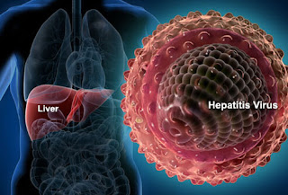
hepatitis virus

Society has been looking at HIV as the most dangerous virus in the world. No wonder if all the power is deployed to combat the virus. But it turns out hepatitis B is much more dangerous than HIV. The high risk of hepatitis B infection even reached 100 times than the virus causes the aids.

The spread of hepatitis B is increasingly vulnerable in endemic countries. Recorded World Health Organization (WHO), from 6 billion of the earth's population, 2 billion of them or as much as 30 percent positive infected with hepatitis B. And based on the map of the spread of hepatitis B made by WHO also, Indonesia including countries with moderate to high infections.

The rate of hepatitis B infection is stated low if only diidap less than 2 percent of the population. Being between 2-8 percent and if more than 8 percent, it means the spread of hepatitis B virus somewhere is high.

"In Indonesia, for areas in Java Island infected with Hepatitis B below 8 percent, but for outside Java reaches more than 8 percent.That is why Indonesia entered the country endemi hepatitis B is high to high," said the expert in internal medicine and consultants digestion and liver From RSUP Kariadi Semarang, Dr. Hery Djagat Purnomo SpPD.

To prove more realistic about the spread of hepatitis B in Indonesia, in 2007 the Ministry of Health conducted basic health research involving 10 thousand samples. As a result, 9.4 percent of the samples were positively infected with hepatitis B.

Given the dangers posed by hepatitis B, Indonesia and Brazil also pioneered the hepatitis B infection care movement so it was determined that July 28 is the day of hepatitis B worldwide. It is expected that public awareness of the danger of this disease also increases.

So, what makes this virus so dangerous? Dr Hery Djagat reveals among others because the distribution is very high, while we do not know anyone around us who suffer from hepatitis B. Patients themselves are often not aware that they are already infected. This is very dangerous because the worst effects of this virus can lead to liver cancer.

Given the heart is one of the most vital organs possessed by humans with the primary function of filtering toxins in the body, you can imagine what happens if the heart is damaged.

Transmission or transmission of hepatitis itself is divided into two vertical transmission and horizontal transmission. Vertical transmission occurs when a mother is suffering from hepatitis B so that at the time of delivery it is very likely to be transmitted to the child. "This is the most common type of transmission, so in Indonesia, every newborn baby should get anti-hepatitis injections," explains Dr. Hery Djagat.

While horizontal transmission is the transmission that occurs between people with people through the media such as blood transfusion, syringes used more than one person, and sexual intercourse.

Based on the amount of viral load and the ability to channel, the highest risk is blood transmission either transfusion, syringe or open wound. The moderate is the transmission through semen / vagina (during intercourse) and saliva or saliva.


What also has the potential to transmit hepatitis B is sweat, milk and tears. It's just that the potential is very small. "For that, in conducting blood transfusion, the use of needles in need of sex especially out of wedlock and others need to be careful," said Dr. Hery Djagat. Msi

KIDNEY STONE: How is the examination, treatment and prevention?


Examination and diagnosis
If your doctor suspects you have Kidney Stones, you may undergo diagnostic tests and procedures, such as:

• Blood tests. Blood tests can show the amount of calcium or uric acid in your blood. Blood test results help monitor your kidney health and may cause your doctor to check other medical conditions.
• A 24-hour urine test may indicate that you are excreting too much rock-forming minerals or too few rock blocks. For this test, your doctor may ask you to do urine collection twice, for two consecutive days.
• Imaging. An imaging test may show Kidney Stones in your urinary tract. Examinations such as plain abdominal xfoto, as well as abdominal CT scan are required.
• Other imaging options include ultrasound, noninvasive tests, and intravenous urography, requiring the injection of dye into the blood through the arm vein and taking X-rays (intravenous pyelogram) or obtaining a CT (CT urogram) imaging CT image through the kidney and the bladder Urine.
• Stone analysis. You may be asked to urinate through a strainer to catch rocks flowing with urine. Laboratory analysis will reveal the composition of your Kidney Stone. Your doctor uses this information to find out what is causing your Kidney Stones and set up a plan to prevent more Kidney Stones

Treatment
Small stones with minimal symptoms
Most Kidney Stones do not require invasive treatment. Things that can be done with small stones are:

• Minimize water. Drinking as much as 2 to 3 liters per day can help clear your urinary tract system.
•          Painkiller. Small stones that pass through the urinary tract can cause pain and discomfort. To reduce pain, your doctor may recommend pain relievers such as ibuprofen, acetaminophen.
• Medical therapy. Your doctor may give you medicine to help get Kidney Stones out through the urinary tract. This type of drug is known as an alpha blocker, and it works by relaxing the muscles in the ureters, helping the Kidney Stone to pass through the urinary tract faster and with less pain.

The stones are big and that cause the symptoms
Kidney stones that can not be treated with conservative measures (either because they are too large to pass by themselves or because of bleeding, kidney damage or ongoing urinary tract infections) require more extensive treatment. Procedures may include:
• Using sound waves to break rocks. For certain Kidney Stones, doctors recommend a procedure called extracorporeal shock lithotripsy (ESWL).
• ESWL uses sound waves to create strong vibrations (shock waves) that can break the stone into small sizes and can come out with urine. The procedure lasts about 45 to 60 minutes and can cause moderate pain, so you may be under sedation or mild anesthesia to make you comfortable.
• ESWL can cause blood in the urine, bruises in the back or abdomen, bleeds around the kidneys and other organs adjacent, and discomfort as the stone fragments pass through the urinary tract.
• Surgery is performed to remove the stone with a very large size in the kidney. A procedure called percutaneous nephrolithotomy involves surgery to remove Kidney Stones by using a telescope and a small instrument inserted through a small incision in the back.
• You will receive general anesthesia during surgery and be hospitalized for one to two days when you recover. Your doctor may recommend this surgery if ESWL is not successful.
• Uses urethroscope to remove stones. To remove the smaller stones in your ureters or kidneys, your doctor may use a urethroscope equipped with a camera through your urethra and bladder.
Once the stone location can be identified, a special tool can pick up a stone or break it into pieces that will flow in your urine. Your doctor can then place a "stent" in the ureter to reduce swelling and improve healing. You may need general or local anesthesia during this procedure.
• Parathyroid gland surgery. Some calcium phosphate stones are caused by an overactive parathyroid gland, located at the four corners of your thyroid gland, just below the Adam's apple. If these glands produce too much parathyroid hormone (hyperparathyroidism), your calcium levels can become too high and Kidney Stones can form as a result.
• Hyperparathyroidism sometimes occurs when small and benign tumors form in one of your parathyroid glands or you develop other conditions that cause these glands to produce more parathyroid hormone. Releasing the growth of the glands will stop the formation of Kidney Stones. Or your doctor may suggest a treatment condition that causes your parathyroid glands to produce too many hormones.
Kidney stone prevention

Lifestyle changes
You can reduce your risk of Kidney Stones if you:
• Drink plenty of water. For people with a history of Kidney Stones, doctors usually recommend urination of 2.5 liters of urine a day. Your doctor may ask you to measure your urine results to ensure that you drink enough water.
• If you live in a hot, dry climate or exercise frequently, you may need to drink more water to produce enough urine. If your urine is light and clear, you may drink enough water.
• Eat fewer foods that contain more oxalates. If you tend to build calcium oxalate stones, your doctor may recommend limiting foods that contain lots of oxalate, such as beets, spinach, sweet potatoes, beans, tea, chocolate and soy products.
• Choose a diet low in salt and animal protein. Reduce the amount of salt you eat and choose non-animal protein sources, such as peas. Consider using a salt substitute.
• Continue to consume foods rich in calcium, but be careful with calcium supplements. Calcium in the diet has no effect on the risk of Kidney Stones. Continue to consume foods rich in calcium unless your doctor suggests otherwise. Ask your doctor before taking calcium supplements, as this is associated with an increased risk of Kidney Stones. You can reduce the risk by taking supplements with food. A low calcium diet can improve the formation of Kidney Stones in some people.
• Ask your doctor to get a referral to a dietitian who can help you develop a meal plan that reduces your Kidney Stone risk.

Medicines used
Medicines can control the amount of minerals and acids in your urine and may help in people who make up some kind of stone. The type of medicine your doctor prescribes will depend on the type of Kidney Stone you have. Here are some examples:
• Calcium stones To help prevent calcium stone formation, your doctor may prescribe thiazid diuretics or phosphate-containing preparations.
• Uric acid stones. Your doctor may prescribe allopurinol to reduce uric acid levels in the blood and urine and medications to keep your alkaline urine. In some cases, allopurinol and alkaline substances can dissolve uric acid stones.
• Struvite stones To prevent struvite stones, your doctor may suggest strategies to keep your urine free from the bacteria that cause the infection. Long-term use of antibiotics in small doses can help achieve this goal. For example, your doctor may recommend antibiotics before and for a while after surgery to treat your Kidney Stones.

• Stone cystine. Stone cystine can be difficult to treat. Your doctor may suggest that you drink more fluids so you produce more urine. If that alone does not help, your doctor may also prescribe drugs that reduce the amount of cystine in your urine.

DIABETIC NEPHROPATHY: How is the treatment and prevention?

Treatment of Diabetic Nephropathy The first step in treating diabetic nephropathy is treating diabetes and, if necessary, treat...

Total Pageviews